
-
ULTIMATE SENSORS Inc.
P.O. Box 4243 Stony Brook,
NY 11790, USA - +1 (516) 901 – 7471
- mig@usensors.com
Fluorescence Imaging
Development of fluorescent imaging system for neuro-surgical microscopes providing an enhanced image of fluorescing tumor tissue overlapped with the surgery operation field image visible by the surgeon’s eye throught the ocular lens. The system is based on time gating of flurescence image registered by a black-white CD camera from the visible image, the fluorescence image processing and enhancement, and following insertion of the enhanced and artificially colored image to the optical path of microscope. (Customer: NIEC Inc.)
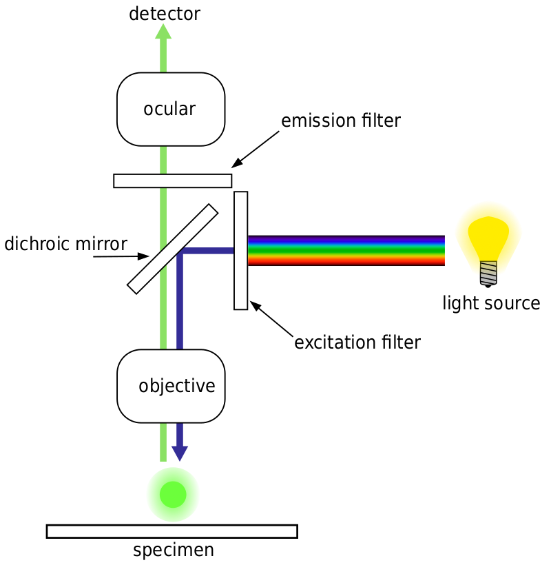
Fig. 1. Lightpath of an epifluorescence microscope. Blue excitation light is reflected by a dichroic mirror filter and excites the sample. The emitted green fluorescence passes through (is transmitted) by the mirror and is detected by a scientific camera. As both the blue excitation and green fluorescence light pass through the objective, it is known as epifluorescent.
Autofluorescence
Some structures, biological organisms, and general microscopy samples can naturally exhibit fluorescence, known as autofluorescence. This is different from fluorescence from labeled samples, but it often shares similar wavelengths, meaning that autofluorescence microscopy samples can obscure artificially added fluorescence and interferes with detection, reducing signal. It is important to know if your samples exhibit autofluorescence, as this will affect any fluorescence imaging done unless specific wavelengths are used to avoid it.
Common examples of autofluorescent objects are mitochondria, lysosomes, collagen and some amino acids such as tryptophan, tyrosine, and phenylalanine. Most notably, autofluorescence is common in plants due to their use of chlorophyll and other fluorescent molecules like lignins and carotenes. Fig.2 shows the different colors of autofluorescence from an unlabeled Scots pine sample.
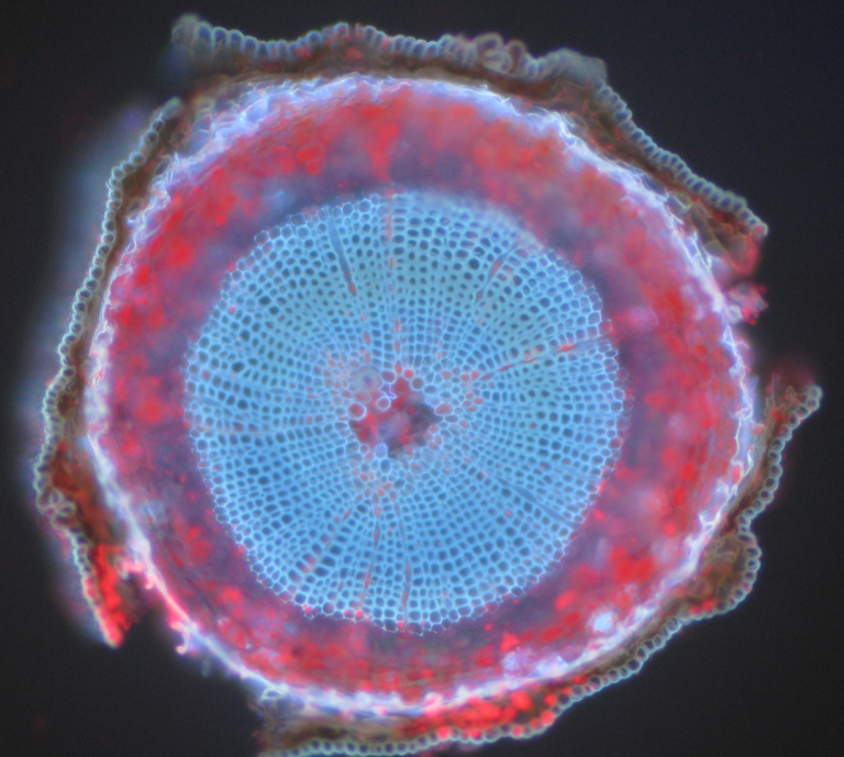
Figure 2: The autofluorescence of a Scots pine sample. This is a composite image (red and blue) from a cross-section of a six-week-old Scots pine seedling, demonstrating autofluorescence in different wavelengths.
Summary
Since the introduction of the first fluorescent dye, fluorescence microscopy has been a heavily utilized tool to visualize cells and cellular structures with higher specificity than traditional brightfield microscopy techniques. Researchers can manipulate the structure, optical properties, and probe of interest in fluorescence experiments to obtain relevant data. Such flexibility has allowed fluorescence microscopy to be included in many life science experiments.
Depending on the type of sample and fluorophore, a scientific camera should be carefully selected to achieve the best imaging results.